
Die Diagnostik histomorphologischer Veränderungen bei Muskelatrophie und Sarkopenie.
DOI: 10.36210/BERMEDJ/epub01062026
Abstract
Sarcopenia refers to the age- or disease-related loss of muscle mass, muscle strength, and muscle function. Morphological changes manifest as a decrease in muscle mass (Cruz-Jentoft 2019), a reduction in muscle fiber count (Lexell 1988), and myosteatosis (Goodpaster 2001). Histological changes associated with sarcopenia include the atrophy of type II muscle fibers (Lexell 1988), the loss of satellite cells (Verdijk 2007), and increased collagen deposition (Narici & Maffulli 2010). Vascular impairment of capillary density is discussed as a potential pathophysiological cause of sarcopenia (Kellum 2020).
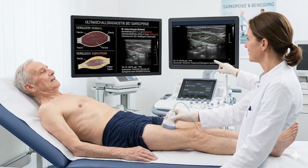
To detect sarcopenic changes, muscle elastography measures the mechanical properties (stiffness/elasticity) of tissues. Sarcopenic muscles are often less stiff, a characteristic attributable to reduced muscle mass (Eby 2015). Fibrosis and fat infiltration give rise to areas of varying stiffness (Akagi 2015).
We examined three groups—each comprising 9 subjects—using muscle elastography (SWE, STE, STQ), performing 24 measurements per subject: 9 healthy volunteers with normal muscle function, 9 patients with muscle atrophy secondary to critical illness polyneuropathy (CIP), and 9 patients with muscle atrophy secondary to spinal cord injury (SCI). The mean BMI was 23.1 in the volunteer group, 19.0 in the CIP group, and 17.5 in the SCI group—figures that may suggest a reduction in muscle mass.
Across the study groups, elastography values ??for the parameters Cs, E, and G differed significantly between the volunteer group and the SCI group (E = 16.9 kPa vs. 11.93 kPa). In the CIP group (E = 14.3 kPa), no significant differences were observed compared to the subject group (E = 16.9 kPa) or the QS group (E = 11.9 kPa). With regard to the reference muscle group, there were no significant differences in shear wave propagation velocity among the study groups.
A key advantage of muscle elastography in a clinical context is its rapid, real-time diagnostic capability. The results are immediately available and provide quantitative measurements in the form of precise tissue stiffness values. Thus, elastography can be utilized as a complementary method for assessing muscle mass and for the early detection of sarcopenia.
Downloads
Published
Versions
- 2026-06-03 (2)
- 2026-06-01 (1)
How to Cite
Issue
Section
License
Copyright (c) 2026 Olaf Schedler; Zdenek Mejzlik, Michal Pobijak, Michael Adamaszek, Wolfgang Laube

This work is licensed under a Creative Commons Attribution-NonCommercial-NoDerivatives 4.0 International License.

